50y/M follow up case.
I HAVE BEEN GIVEN THIS CASE TO SOLVE IN AN ATTEMPT TO UNDERSTAND THE TOPIC OF ''PATIENT CLINICAL DATA ANALYSIS'' TO DEVELOP MY COMPETENCY IN READING AND COMPREHENDING CLINICAL DATA INCLUDING HISTORY,CLINICAL FINDINGS,INVESTIGATIONS AND COME UP WITH A DIAGNOSIS AND TREATMENT PLAN.
A 50 year old male patient is on maintainance haemodialysis since 10 months.
HOPI: patient was apparently asymtomatic 4yrs back and then developed shortness of breath (on and off), pedal oedema (pitting type).Later diagnosed as chronic renal failure and underwent dialysis twice weekly for about 10 months.
6 years back he met with an accident. His right leg got fractured and it took nearly 1 year to heal for which he used few medication continuosly for 1 year.
PAST HISTORY: k/c/o HTN since 1 year.
Not a K/C/O of DM, thyroid disorders, TB
PERSONAL HISTORY:He follows a mixed diet. Appetite -Normal, Bladder movements-normal,
Bowel movements-constipation since few weeks. Sleep- decreased.consumes alcohol regularly(90ml ) and stoped consuming1 year back.
He is a farmer and stopped working since 1 year.
FAMILY HISTORY: No significant family history.
DRUG HISTORY: No known drug allegies and patient uses Nicardia 10 mg.
General examination : patient is conscious ,coherrent, co operative and well oriented to his surroundings.he is poorly built and nourished.no pallor ,no cyanosis, no icterus, no lymphadenopathy. bilateral pedal edema is seen and is of pitting type
Vitals:. Temperature: afebrile. Pulse rate: 98 beats / min. Respiratory rate: 19cycles / min. Bp: 190/100. Spo2: 99
Systemic examination:
Cvs: bilaterally symmetric chest wall .no precordial bulge .no thrills and no murmurs.
S1& S2 heard.
Respiratory system: no dyspnoea, no wheeze
Position of trachea- central, no adventious sounds heard
CNS: patient is normal and concious .reflexs are normal.
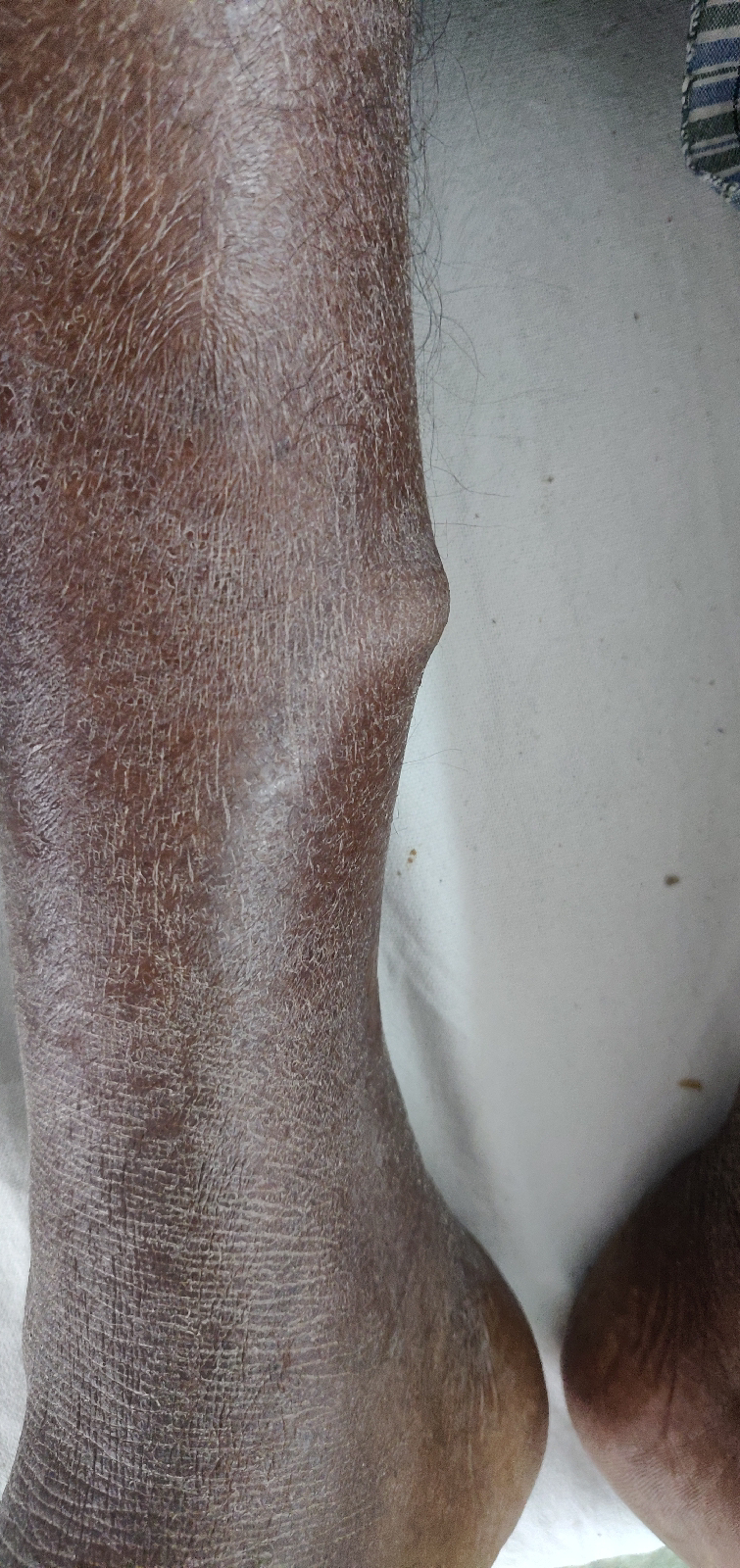
CLINICAL IMAGES:


Investigations: 31/1/22
RFT: urea-157. Cr.10.2. UA-9.8
USG- Rt Grade 3 RPD
Lt grade 2 RPD
2D ECHO- trivial TR+ /AR+, no MR.
Good LV systolic function.
Diastolic dysfunction (+)
ECG:

2/2/22:
LFT:
T.b-0.9. D.B- 0.2. SGOT-17. SGPT-15. ALKP- 504. TP-5.6. ALB-3.6. A/G RATIO-1.80
RFT: U-178. CR-10.2. U A-9.0. CALCIUM-9.4. P-4.5. Na-140. K-4.7. Cl-102
S.iron 78
RBS- 70
CUE: ALB++. SUGARS-TRACE. RBC's, CRYSTALS, CASTS-NIL
HAEMOGRAM
HB-5.8. TLC-7400. LYMPHOCYTES -13
PCV-17.4. RBC COUNT-2.01. PLT-1.20.
NORMOCYTIC NORMOCHROMIC ANEMIA WITH THROMBOCYTOPENIA.
PROVISIONAL DIAGNOSIS:
NSAID ASSOCIATED RENAL IMPAIRMENT.
TREATMENT:
(1) TAB.LASIX 40MG PO/BD
(2) TAB.NICARDIA 10 MG PO/BD
(3)TAB.NODOSIS 500MG PO/BD
(4)TAB.OROFER -XT PO/OD
(5)TAB.SHELCAL-CT PO/OD
(6)INJ.ERYTHROPIEOTIN 4000IU S/C ONCE WEEKLY
(7) SALT AND FLUID RESTRICTION.
COURSE IN HOSPITAL:
Patient has intermittent fever high grade on & off and neck pain since 1 month and neck stiffness with restriction neck movements since 10 days. Xray C-spine was taken which shows no bony abnormalities conservative management was done to relieve the pain. But patient symptoms didn't subsided. Mri C spine with whole spine screening was done. Which shows


SIMILAR CASE REPORT-
A 65–year–old man on regular hemodialysis for long–standing chronic renal failure sought treatment after 2 days of progressively worsening neck pain. The patient described a febrile illness in the days preceding his admission. Examination of his peripheral nervous system was normal other than that he exhibited Lhermitte's sign.4 Based on this history, spinal sepsis was suspected. Blood was drawn for culture and sensitivity and urgent magnetic resonance imaging (MRI) was arranged. Gram–positive cocci were detected in the blood, and empirical therapy with vancomycin, flucloxacillin, and rifampicin was started. MRI of the craniovertebral junction showed an epidural mass around the odontoid process causing cervicomedullary compression and an abnormal hypointensity in the area on the right side of C1 . Subsequently, the bacteremia was confirmed to be due to Staphylococcus aureus. Day 3 of admission, the patient developed signs of respiratory distress and carbon dioxide retention.


Comments
Post a Comment